Tissue microarrays (TMA) are tools to study disease biomarkers in well-defined patient populations. Our organization supports basic research into fatty liver disease, offering high quality test systems and publishing research to help improve the availability of representative tissue samples and other relevant test systems (e.g., human liver microsomes and S9 from NASH donors). We developed liver tissue microarrays for analysis of the progression of fatty liver disease in non-alcoholic steatohepatitis (NASH) and alcoholic steatohepatitis subjects.

Tissue Microarray (TMA) Offerings for Liver Disease & Other Research
Tissue sourced from our Research Biobank is mounted unstained with no overlay in 3mm diameter cores which are 4µm thick. Staining options (e.g., H&E images) are available upon request for convenience. All arrays come with information about donors’ gender, age, ethnicity, and BMI and datasheets can be customized for additional characteristics, such as alcohol use and diabetes, for more specific comparison (see all inventory and customization options here).
TMA Preparation and Disease-Specific Research Applications
Tissue microarrays are used in a higher-throughput mode, alleviating variability associated with individual conventional lot-to-lot comparison which can have inconsistencies skewing results and are less efficient. TMAs can be used for a wide variety of studies, including biomarker analysis and protein and gene profiling. Additionally, the uses of tissue arrays cater to values in efficiency and quality. TMAs boast “substantial benefits over standard techniques and represents a significant advancement in molecular pathology.” For example, TMA technology reduces laboratory work, offers a high level of experimental uniformity and provides a judicious use of precious tissue.1
Research areas which may benefit from Research Biobank tissue microarrays include:
- Non-alcoholic steatohepatitis (NASH)
- Non-alcoholic fatty liver disease (NAFLD)
- Alcoholic steatohepatitis
- Liver fibrosis, cirrhosis
- Diabetes
Tissue Preparation
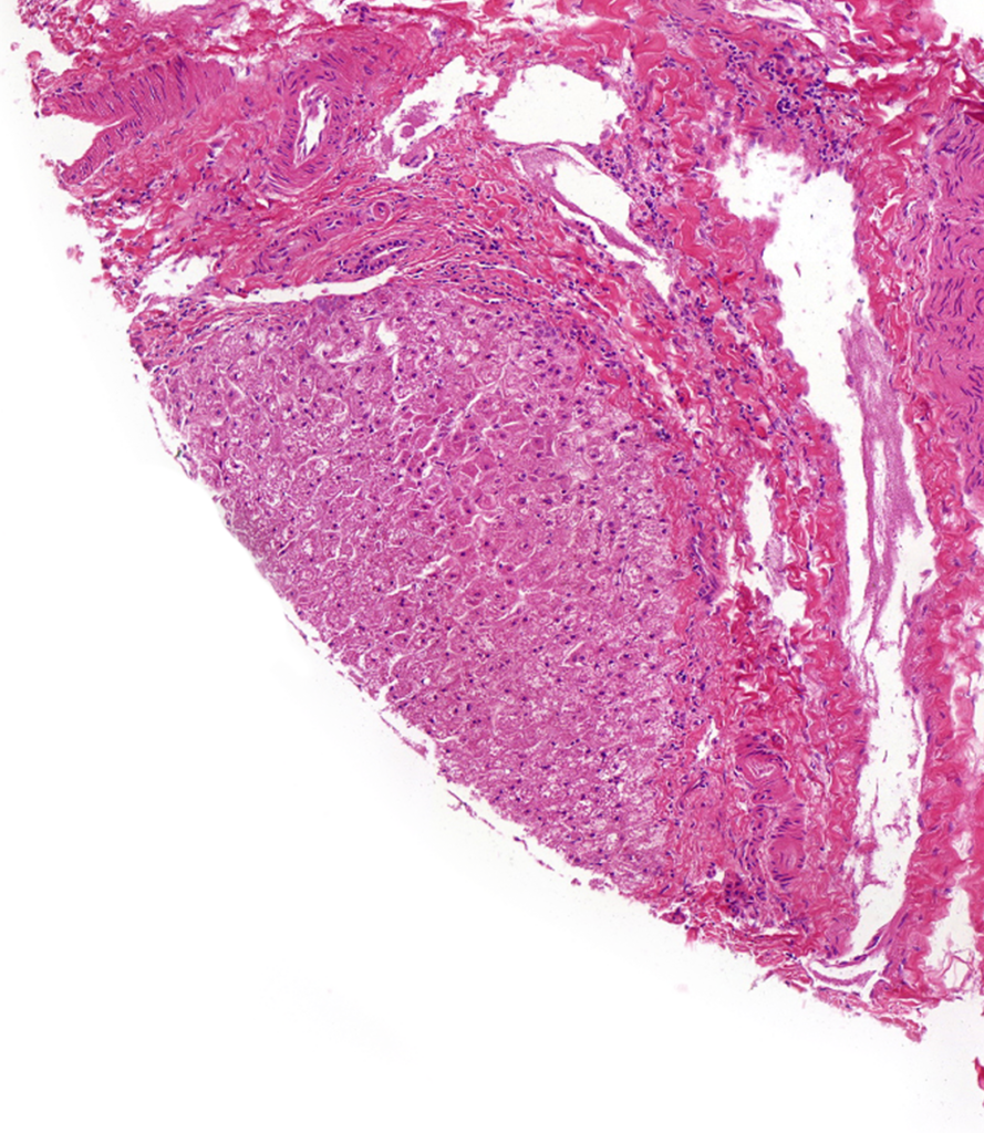
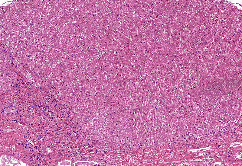
The placement of targeted disease tissues and appropriate controls in the same array facilitates comparative analysis of biomarker distribution. Tissue donors’ data, including BMI and macrovesicular fat content, accompany each array. Our arrays are built with donated Research Biobank tissues, which were collected and preserved with the initial intent for human transplantation. The tissues are mounted on a typical microscope slide and are not stained or covered/overlaid unless otherwise requested. The microarrays are cut in small batches and stored at 4° C in atmosphere depleted of oxygen. The images below of hematoxylin/eosin (H&E) and Masson’s trichrome-stained arrays illustrate histology and fibrosis on 3 mm diameter cores:

Pediatric Tissue Microarrays
Pediatric liver disease affects hundreds of thousands of families around the world. “Non-alcoholic fatty liver disease (NAFLD) is a major cause of chronic liver disease, and is the most common form of chronic liver disease in many industrialized countries. In the United States, there are over 64 million people with NAFLD, with annual direct medical costs of about $103 billion ($1,613 per patient).”2 NAFLD is the most common cause of chronic liver disease in the United States, with nearly ¼ of patients progressing to NASH3. The need to explore therapeutic avenues towards NAFLD is on the rise, necessitating research specific to pediatric liver function and drug response.

We have recently introduced pediatric tissue options for researchers in this field. The pediatric hepatic tissue array is comprised of 25 samples representing a range of disease status with an additional 5 controls. Read more about our pediatric offerings and applications in our recent blog post.
How we characterize normal vs diseased tissue
Tissue classifications reflect only major features of fatty liver disease, these tissues may have characteristics of other liver disease(s).
Science related to TMA use in NASH, NAFLD, and other liver disease:
Webinar: Drug Metabolizing Enzymes and Transporters in NASH
Poster: CYP1A2 Enzyme Activity and Protein Abundance In Normal And Diseased Pediatric Livers
Poster: Patterns of Hyaluronan Deposition in Normal and Diseased Pediatric Livers
Poster: Microsomal Cytochrome P450 Enzyme Activities in Nonalcoholic Steatohepatitis Livers
References
- Quagliata L, Schlageter M, Quintavalle C, Tornillo L, Terracciano LM. Identification of New Players in Hepatocarcinogenesis: Limits and Opportunities of Using Tissue Microarray (TMA). Microarrays (Basel). 2014;3(2):91-102. Published 2014 Apr 15. doi:10.3390/microarrays3020091
- Tungland B, “Oral Dysbiosis and Periodontal Disease: Effects on Systemic Physiology and in Metabolic Diseases, and Effects of Various Therapeutic Strategies.” Human Microbiota in Health and Disease: from Pathogenesis to Therapy. Elsevier, 2018.
- Perumpail, Brandon J. “Clinical Epidemiology and Disease Burden of Nonalcoholic Fatty Liver Disease.” World Journal of Gastroenterology, vol. 23, no. 47, 21 Dec. 2017, pp. 8263–8267., doi:10.3748/wjg.v23.i47.8263.

