Mitochondria play an important role in maintaining life activity, including adenosine triphosphate (ATP) production. We offer a Mitochondrial Toxicity study developed by our partners at the Drug Development Solutions Center to assist you in your drug’s hepatotoxicity / drug induced liver injury (DILI) risk assessment.

Mitochondrial Toxicity Assay to Evaluate DILI Risk of Your Drug Candidate
You can now request quotes for our research services on BioIVT.com!
Whether you need a single assay or a complete ADME program, BioIVT’s experts will help design and implement the appropriate studies for your drug and research objectives. View BioIVT’s comprehensive portfolio of ADME research services.
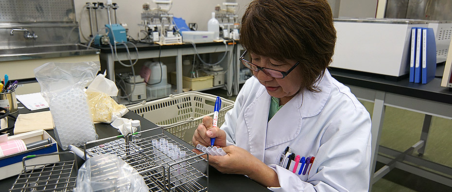
In this contracted in vitro study, human hepatoma-derived HepG2 cells are cultured with the test compound in two different media to determine whether or not there a decrease in ATP generation, a useful endpoint to assess a drug’s effect on mitochondrial activity.
Data supporting the reliability of this assay can be found in our 2016 scientific poster, “A simple separated evaluation method for mitochondrial toxicity using Crabtree effect” presented at the JSSX annual conference by leading scientists at the Drug Development Solutions Center.
Study Design to Determine Mitochondrial Toxicity
Experimental Evaluation in Glucose vs Galactose
Highly proliferative cells, such as HepG2, are adapted to anaerobic conditions and respond best to high-glucose media. As a result, these cells favor glycolysis over oxidative phosphorylation for producing ATP, a process which does not necessitate normal mitochondrial function. This is known as the Crabtree Effect. Current methodologies in toxicology still use immortalized cell lines like HepG2, overcoming the Crabtree Effect by using galactose in cell media, forcing the cells to generate ATP via oxidative phosphorylation, therefore making it possible to observe downstream effects of mitochondrial toxicity.
Comparing effects on cells between glucose- and galactose-rich media can identify or rule out cytotoxicity caused specifically by mitochondrial impairment.

Long vs Short Term Incubation
We can offer short- or long-term exposure options for the assay, depending on your interest. You may choose a short-term experiment if you are interested in:
- ATP synthesis inhibition
- Respiration chain inhibition (MRC)
- Uncoupling
Short term experiments are performed in triplicate, measuring ATP production after 1 day of incubation with multiple concentrations of test article versus positive control (oligomycin).
You may opt for the long-term exposure option if you are interested in ribosomal inhibition. Long-term incubations can range from 6 to 14 days. This experiment is performed in triplicate, measuring ATP at pre-determined time points over the course of the incubation. The exposure media is changed every 2-3 days to maintain culture conditions for the cells, and the positive control used is chloramphenicol. Multiple concentrations of test article can be included to establish concentration-dependence.
ATP Measurement
After exposure to the drug for a specified period of time, the ATP production amount is measured using CellTiter-Glo Luminescent Cell Viability Assay, and the IC50 is calculated from the ATP production decrease rate.
A ratio (%) is then calculated, comparing ATP emission intensity in the presence of test substance or positive control versus absence of test substance or positive control. This ratio is then used to calculate EC50 value, representing the relationship between concentration and the ATP amount ratio (%).
Results
Following conclusion of the experiment, the sponsor receives a data report of:
- ATP amount ratio (%)
- EC50 (µmol/L or nmol/L)
- Fold change
Understanding DILI Risk: Mitochondrial Toxicity & BSEP Inhibition
As scientific understanding of DILI grows and more instances of mild to severe liver damage are documented, patterns have emerged that indicate multiple predictable drug-related factors that could put a patient at higher risk for cholestasis. About 80% of the boxed warning compounds reported by the FDA shows mitochondrial toxicity, thus it has been widely regarded as a standard study for toxicity screening of drug candidates in Discovery & Development.

A related correlation with growing experimental support proposes a connection between BSEP inhibition and mitochondrial toxicity in predicting acute DILI in the clinic. In the incidence of mitochondrial toxicity, as mitochondria stop working to produce ATP, many components of the cell reliant on ATP—including ATP-binding cassette (ABC) transporters—don’t have the energy source to function. The bile salt export pump (BSEP) is an ABC transporter found in high concentrations in the walls of hepatocytes and function to excrete bile acid from inside the cell. With impaired BSEP function, accumulation of toxic bile acids could lead to apoptosis of hepatocytes, causing liver injury (cholestasis).
If a drug has potential both to inhibit BSEP and impair mitochondrial function through toxicity, it could lead to bile acid-dependent hepatotoxicity more than if it is affected by one factor by itself, leading to a higher risk of severe DILI.
This theory is supported by a 2014 study which examined 72 different xenobiotic compounds characterized by mitochondrial toxicity, apparent BSEP inhibition potential, or both, and correlated to varying degrees of clinical concern. The study found that “drugs with dual potency as mitochondrial and BSEP inhibitors were highly associated with more severe human DILI, more restrictive product safety labeling related to liver injury, and appear more sensitive to the drug exposure (Cmax) where more restrictive labeling occurs” [1].
If you are interested in supplementing your mitochondrial toxicity data with BSEP inhibition studies, we offer in vitro drug transporter inhibition studies as well as a Bile Acid-Dependent Hepatotoxicity assay. We also offer other in vitro assays to assess Hepatotoxicity, including Covalent Binding, Reactive Metabolite Identification, and Acyl-Glucuronide Formation.
References
- Aleo, Michael D., et al. “Human Drug-Induced Liver Injury Severity Is Highly Associated with Dual Inhibition of Liver Mitochondrial Function and Bile Salt Export Pump.” Hepatology, vol. 60, no. 3, 2014, pp. 1015–1022., doi:10.1002/hep.27206.

