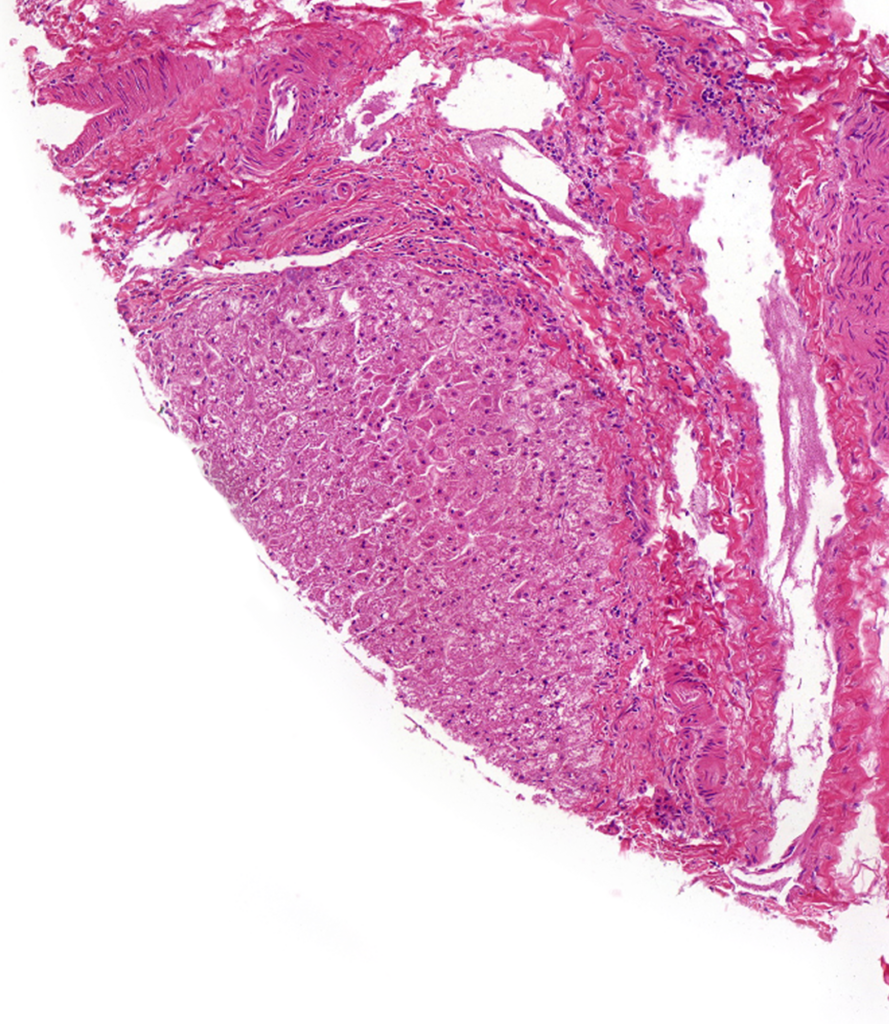
We offer pooled enteric subcellular fractions isolated from enteric (intestinal) tissue from male Beagle dog, demonstrating high enzyme activity levels. Options include matching microsomal, S9, and cytosolic fractions prepared with or without PMSF.

Canine (Dog) Intestine Microsomes, S9 & Cytosol
XenoTech products are now available for purchase at BioIVT.com!
Browse BioIVT’s hepatocytes, subcellular fractions & recombinant enzymes and create an account on bioivt.com to view pricing, generate quotes, and purchase products. Click here for a guide to the new products’ part numbers.
Intestinal microsomes are supplied at a protein concentration of 10 mg/mL in 250 mM sucrose buffer. S9 (4 mg/mL) and cytosolic (4 mg/mL) fractions are supplied in a homogenization buffer with or without PMSF– find out more about elution and sample preparation on page 80 of our Product Tech Guide.
Utilizing Canine Intestinal Subcellular Fractions in Preclinical Research
Use of pooled intestinal subcellular fractions as a test system is highly recommended due to the importance of intestinal enzymes in the first-pass metabolism of many orally ingested xenobiotics, including cytochrome P450 (CYP), UDP-glucuronosyltransferase (UGT), and esterase enzymes. Consequences of intestinal biotransformation can be very significant, including toxification by bioactivation and detoxification by aiding in excretion.
Our highly experienced products team is well-practiced in isolating enteric test systems to ensure superior data by using only mature villus tips in the preparation of intestinal subcellular fractions. Other parts of the intestine, including the submucosa, muscularis and serosa, yield low microsome quantities and are discarded in order to ensure the highest quality intestinal subcellular fractions on the market. Our unique preparation methods allow for large lots for consistency between assays and availability of matching S9, microsomes, and cytosol for correlation between studies using different test systems.
Microsomes contain the highest concentration of many important drug-metabolizing enzymes including cytochrome P450 (CYP) and UDP-glucuronosyltransferase (UGT) enzymes. S9 fractions contain a mixture of cytosolic and microsomal esenzymes, which express a wide variety of phase I and phase II enzymes and are recommended for many different PK/ADME studies. Cytosolic fractions contain many unique drug-metabolizing enzymes that are not enriched in microsomes and are a recommended test system for evaluating soluble phase II metabolic reactions of a test compound – especially once reactions have been observed occurred in S9 fractions, as the microsomal enzymes have been significantly reduced from the cytosol fraction allowing for enhanced observation of non-CYP metabolism.
Why are some lots PMSF-free?
Most ester-type drugs are hydrolyzed by esterases bound in the intestinal mucosa1, so esterase activity in intestinal test systems can be useful in understanding a drug candidate’s extrahepatic metabolic profile. Phenylmethylsulfonyl flouride (PMSF), a serine protease inhibitor, is included in buffers used during tissue processing and subcellular fraction because it protects CYP and other important drug-metabolizing enzymes from being broken down by the high levels of naturally occurring proteases found in the intestinal compartment. The compromise to protecting those metabolic activities is that the PMSF also inhibits esterases.
To remedy this, we offer PMSF-free fractions from human, monkey, rat, and dog. The PMSF-free fractions have unimpeded esterase activity, however it comes at the detriment of other metabolic activities due to increased protease liability so we recommend PMSF-free fractions for use in assays investigating esterase interaction specifically. For all assays focused on other enzymes (e.g. CYP, UGT, SULT) we recommend our standard intestinal subcellular fractions prepared with PMSF.
Read more about elution methods and activity characterization in a recent blog post by Senior Manager of Technical Support, Dr. Chris Bohl: Considerations when studying esterase activities in the intestine.
Make sure your microsomes and S9 fractions are properly activated
For best results from assays using microsomes and S9 fractions, we recommend using NADPH regeneration to support high metabolism levels by ensuring that the NADPH levels are not a limiting factor in your incubations. Our preferred system, RapidStartTM, uses an enzymatic reaction that converts NADP to NADPH, which is then used as a cofactor by CYPs and oxidized back to NADP, and the cycle continues ensuring stable NADPH levels throughout the incubation. RapidStartTM NADPH regeneration is easily activated by simply adding water. Our easy-to-use formula is the most convenient and flexible NADPH regenerating system you’ll find, allowing users to easily tailor the system’s capability by simply varying the amount of water added. RapidStartTM is perfect for long- and short-term incubations and supports the function of recombinant enzymes and subcellular fractions, allowing you to achieve the most accurate and reliable data during your in vitro assays.
Don’t see what you need? Other preparations can be made available!
If you require subcellular test systems not available through our standard product offerings, Custom Subcellular Preparations can be easily ordered to meet the specific needs of your study.
Our designated Custom Products team regularly prepares and isolates cells and/or subcellular fractions from more than 45 different species and has capabilities to meet unique needs that are unavailable through most vendors– non-standard rodent strains, farm animals, insects, precarious tissue isolations such as adrenal gland or jejunum. Our specialists can carry out over 40 characterization assays, providing the end user with a good foundation for their DMPK assays. See our Custom Products page for examples of past custom preparations or get in touch with a specialist to find out how we can meet your needs.

References
- Inoue, Michiko, et al. “Species Difference And Characterization Of Intestinal Esterase On The Hydrolizing Activity Of Ester-Type Drugs.” The Japanese Journal of Pharmacology, vol. 29, no. 1, 1979, pp. 9–16., doi:10.1254/jjp.29.9.

